The state-of-the-art laboratory is located at the Research Center of the Institut universitaire de gériatrie de Montréal (CRIUGM), at 4545 Queen-Mary Road in Montreal.
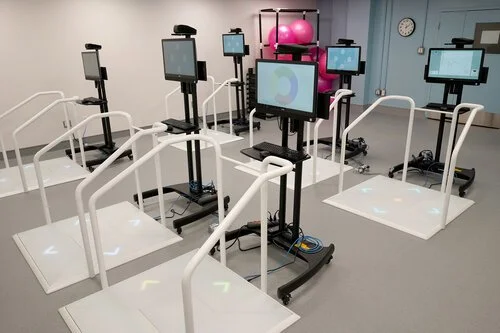
The lab was first established through Canadian Foundation on Innovation (CFI) infrastructure grants of $208K (2007), $245K (2014) and $200K (2018), which ensure access to cutting-edge measuring instruments and equipment. Supported by CRIUGM spaces and utilities, Dr. Dumoulin’s laboratories comprises three evaluation/treatment rooms, an equipment disinfecting/sterilization room, a gym equipped with mirrors, parallel bars and a virtual-reality rehabilitation room with 10 dance mats/computers stations. Other key equipment include three ultrasound scanners with perineal and endo-vaginal 3D/4D probes to assess pelvic floor muscle morphometrics and vascularity, two pelvic floor muscle dynamometers to assess pelvic floor muscle function, two biofeedback/electrical stimulation rehabilitation units for pelvic floor muscle training and 10 virtual reality stations.
Enjoy your virtual visit!
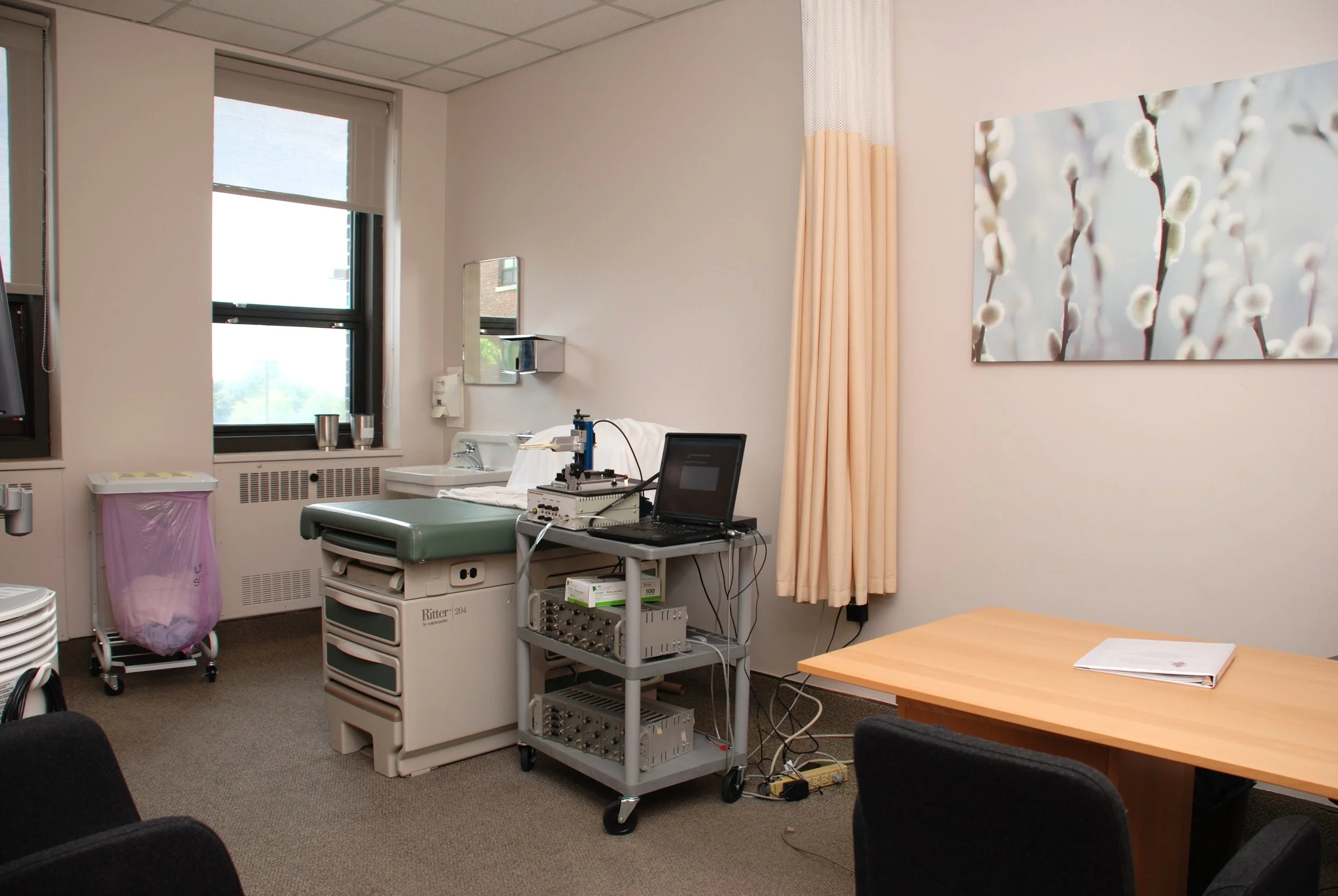
Evaluation rooms

Advanced technologies

In the laboratory, the ultrasound machine is mainly used to make morphometric measurements of the pelvic floor muscles and pelvic organs at rest, during exertion (coughing or pushing) and contraction. This is done by placing a probe on the perineum. The ultrasound device is also used to measure the vascularity of the internal and dorsal pudendal arteries of the clitoris, using the Doppler mode on the device.

Pelvic floor dynamometry measures the force produced by the pelvic floor muscles. A dynamometer looks like a small speculum with two narrow branches (1.5 cm), which are inserted into the vagina at a depth of 5 cm. When the muscles contract, the pressure exerted on the branches of the dynamometer is recorded on a computer and provides information on the tone, strength and endurance of the pelvic floor muscles.

A bladder scan is a non-invasive, portable ultrasound device that measures the volume of urine retained within the bladder. It is a quick, easy and painless tool, that can scan the bladder within a few seconds. It uses high-frequency low-energy sound waves, which are reflected by the bladder tissue. In the laboratory, it is mostly used to objectively assess bladder volume and perform a cough test at a standardized capacity.

The dual-energy X-ray absorptiometry (DEXA) scan uses two low-energy X-ray beams. Since soft tissues and bones have different x-ray attenuation coefficients, using two energies can differentiate the structures and calculate bone density and fat percentage. In our laboratory, it is a useful technology to calculate body fat percentage. Body fat percentage is correlated with stress urinary incontinence because of increased intra-abdominal pressure.
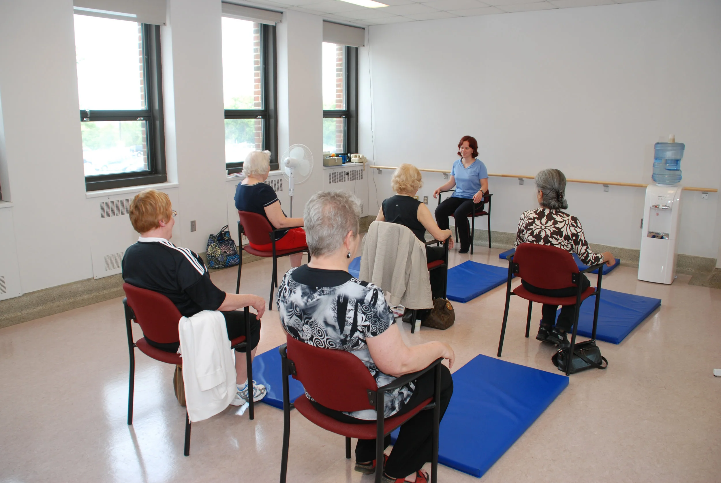
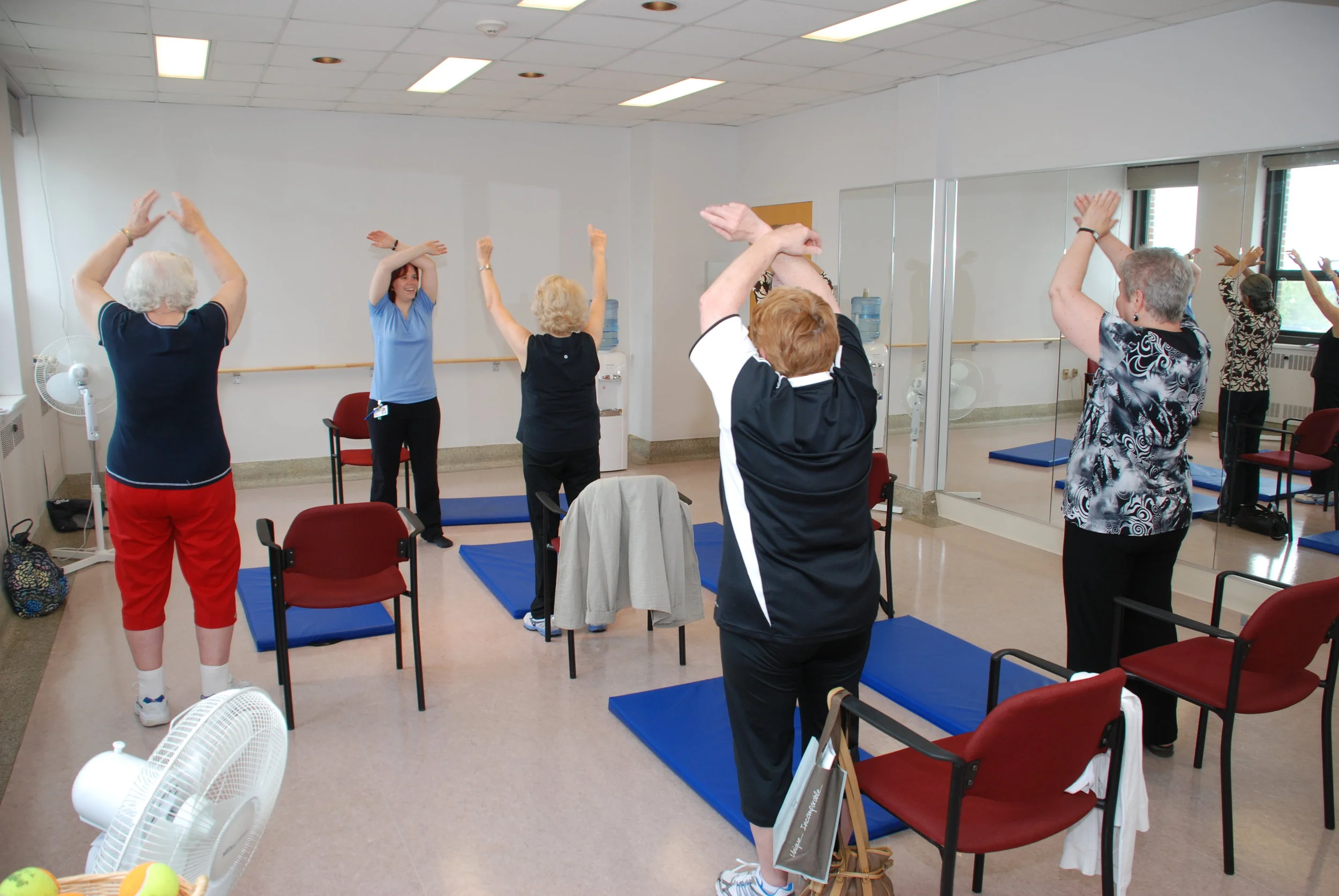
Individual and group treatments

Individual perineal physiotherapy treatments are available for certain projects in our laboratory. They include education on the various urogynecological pathologies of interest and pelvic floor muscle rehabilitation. Pelvic floor muscle rehabilitation involves specific exercises to strengthen and normalize pelvic floor muscle function, and may also include the use of biofeedback (EMG biofeedback) and electroneurostimulation. EMG biofeedback informs the patient about the electrical activity produced by their pelvic floor muscles, while electrostimulation induces contraction of the pelvic floor muscles to facilitate muscle recruitment. Both interventions can be used to improve pelvic floor muscle function and strengthen weak or atrophied muscles.
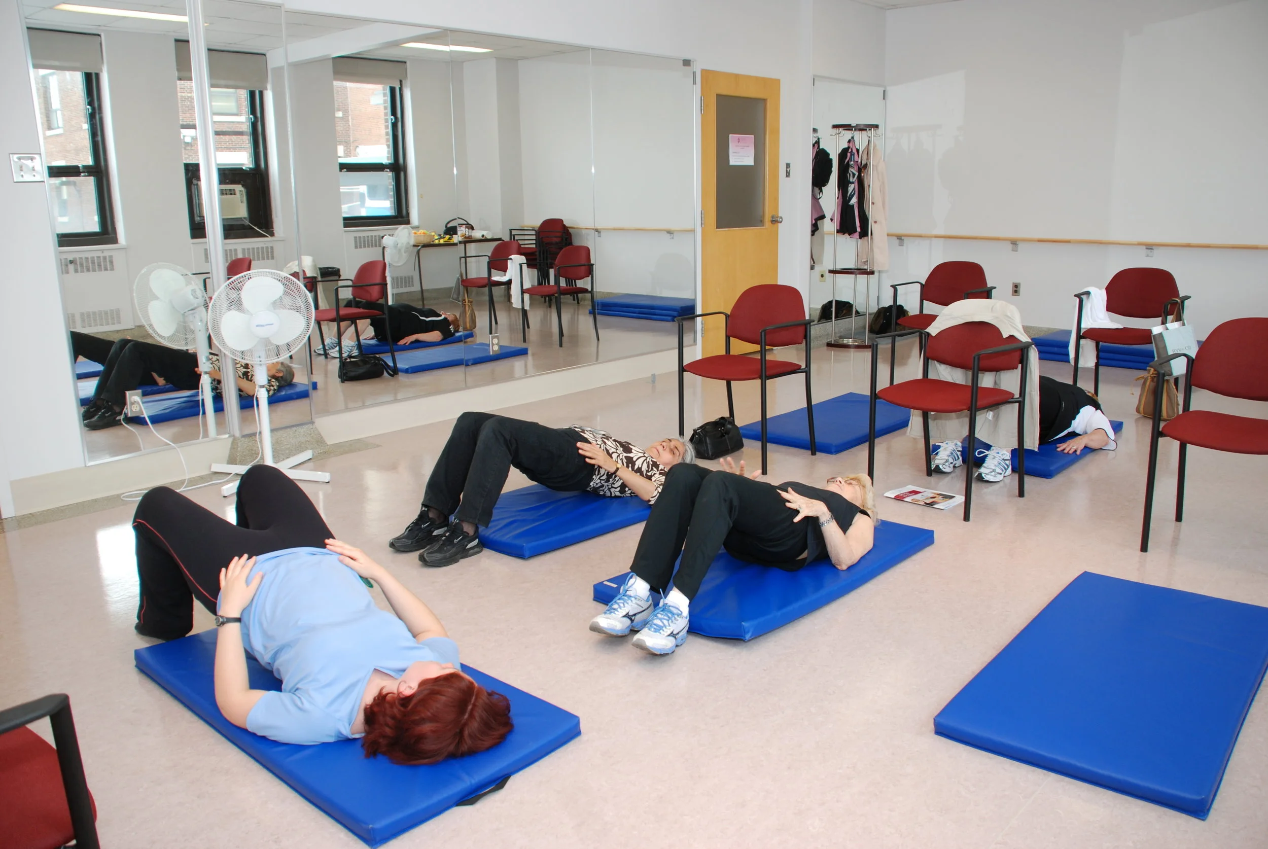
Pelvic floor muscle exercise classes for the treatment of urinary incontinence and other pelvic floor dysfunctions take place in the laboratory’s gym. They include an educational component on relevant urogynecological pathologies and pelvic floor muscle rehabilitation. Pelvic floor muscle rehabilitation includes strengthening, endurance and coordination exercises in different positions: lying down, on all fours (in a table-top position), sitting and standing. Along with these classes, participants receive a home exercise program.
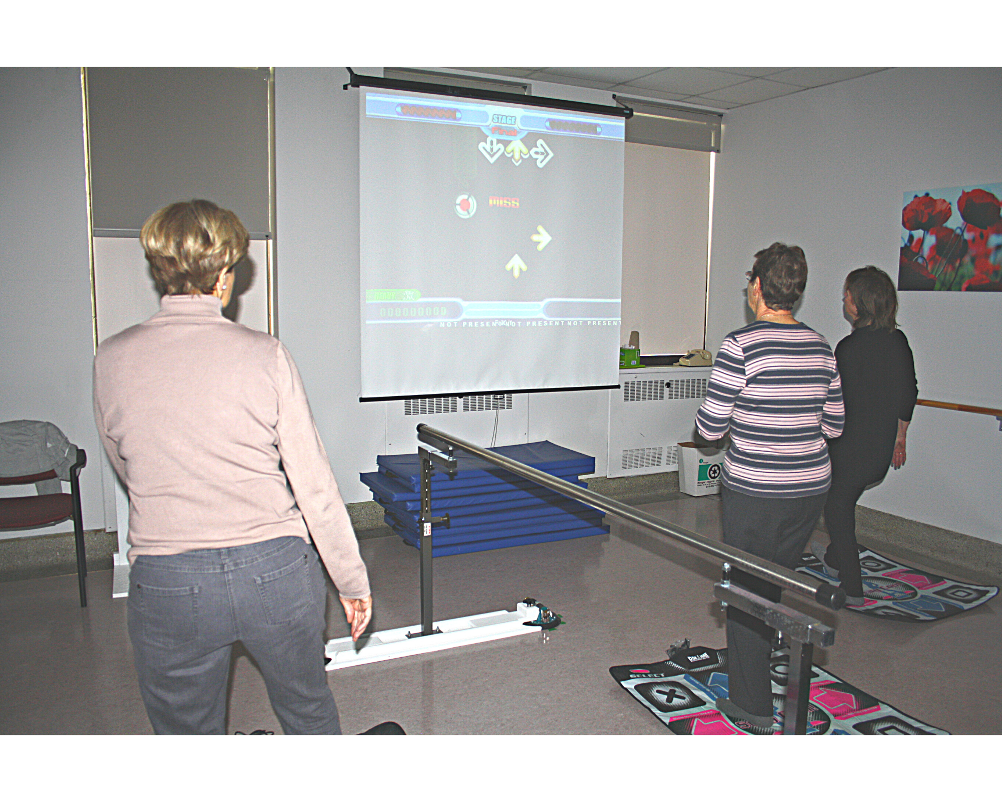
Virtual reality
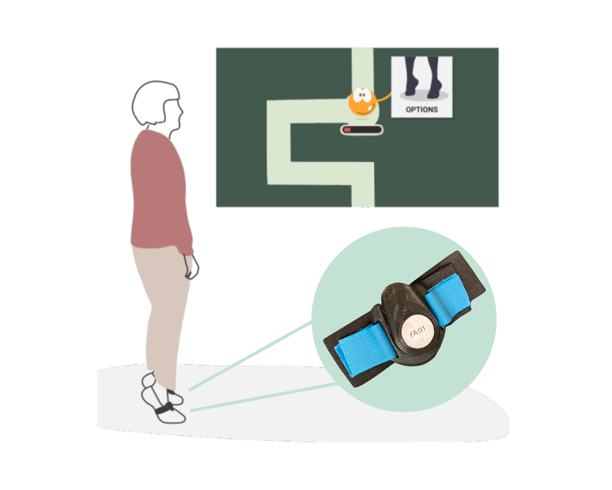
Virtual Reality can be used for pelvic floor muscle rehabilitation. Virtual reality helps participants use technology to learn to activate their pelvic floor muscles while performing functional movements, which resemble everyday activities. Performing pelvic floor muscle contractions while doing another task, such as dancing, requires not only physical effort from the pelvic floor muscles, but also from the legs, as well as a cognitive effort.
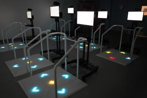
Here are the devices used for virtual reality rehabilitation in our laboratory:
Stepmania is a dance game where the player follows a sequence of steps on a dance mat, while contracting their pelvic floor muscles to the sound of rhythmic music.


2. Dividat's Senso© platform is a training system consisting of interactive games that improve users’ physical and cognitive functions. The platform is equipped with sensors that measure and record various strength and balance parameters of the lower limbs during the games.
3. The VITAAL Exergame is an innovative comprehensive system for geriatric rehabilitation. It uses wearable sensors and a web-based interface to provide evidence-based motor-cognitive and incontinence training with high usability and quick/barrier-free application in the clinic and at home.